Vitreoretina
What does vitreoretinal mean?
Vitreoretinal is a term that refers to the interface between the vitreous body, a gel-like substance filling the space between the lens and the retina in the eye, and the retina itself. Understanding the significance of this term requires delving into the anatomy and physiology of the eye.
The vitreous body, or simply vitreous, is a transparent gel-like substance that occupies about two-thirds of the eye's volume. Composed mainly of water, it also contains collagen fibers and other macromolecules that give it its gel-like consistency. The vitreous plays crucial roles in maintaining the shape of the eye, providing a clear pathway for light to reach the retina, and supporting the delicate structures within the eye.
The retina, on the other hand, is a thin layer of tissue located at the back of the eye. It consists of several layers of cells, including photoreceptor cells (rods and cones) that convert light into electrical signals, and various types of neurons that process and transmit these signals to the brain via the optic nerve.The retina is essential for vision, as it serves as the sensory receptor layer that captures visual information and initiates

the process of visual perception.
The vitreoretinal interface, therefore, refers to the region where the vitreous body comes into contact with the retina. This interface is critical for the overall health and function of the eye. However, various conditions can affect the vitreoretinal interface, leading to vision problems and potential complications.
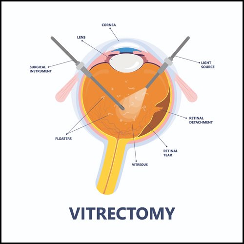
One common condition affecting this interface is vitreoretinal traction, where the vitreous exerts abnormal pulling forces on the retina. This can occur due to age-related changes in the vitreous structure, leading to the formation of tractional bands or adhesions between the vitreous and the retina. Vitreoretinal traction can cause symptoms such as floaters (perceived specks or strands floating in the visual field), flashes of light, and even retinal tears or detachments if the tractional forces are strong enough to cause mechanical damage to the retina.
Another condition involving the vitreoretinal interface is vitreomacular traction (VMT), where there is abnormal adhesion or traction specifically at the macula, the central part of the retina responsible for detailed central vision. VMT can lead to distortion or blurring of central vision, making tasks like reading or driving difficult.
Additionally, diseases such as diabetic retinopathy and age-related macular degeneration can also affect the vitreoretinal interface, leading to vision loss and other complications if left untreated.
the vitreoretinal interface is a crucial anatomical and functional aspect of the eye, where the vitreous body interacts with the retina. Understanding and managing conditions affecting this interface are essential for preserving vision and maintaining eye health.

We are here to help you.
Book An Appointment
I need to schedule an appointment with the eye specialist for a comprehensive eye examination. Please confirm the earliest available slot. Thank you.
Call Now
Please call now to book an appointment with the eye specialist at your earliest convenience. I require a consultation soon. Thank you.

Whatsapp Now
Feel free to send a WhatsApp message now to arrange an appointment with the eye specialist. Urgency is appreciated. Thank you.
What is a vitreoretinal procedure?
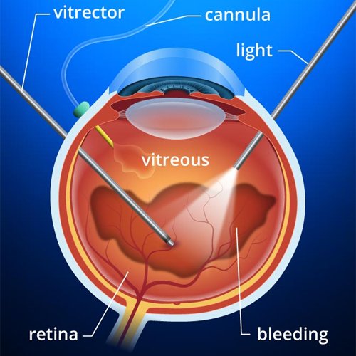
A vitreoretinal procedure refers to a variety of surgical techniques aimed at treating disorders related to the retina and vitreous humor within the eye. These procedures are crucial in managing conditions that can significantly impair vision or lead to blindness if left untreated. The retina is the light-sensitive tissue at the back of the eye responsible for converting light into neural signals that the brain interprets as vision, while the vitreous humor is the clear gel-like substance that fills the space between the lens and the retina.
Vitreoretinal procedures are employed to address a range of conditions, including retinal detachment, diabetic retinopathy, macular holes, epiretinal membranes, and vitreous hemorrhage. Retinal detachment, a medical emergency, occurs when the retina separates from its underlying supportive tissue, which can lead to permanent vision loss. Diabetic retinopathy, a complication of diabetes, involves damage to the retinal blood vessels and can cause bleeding, swelling, and scarring.
Common vitreoretinal procedures include vitrectomy, scleral buckle

surgery, and laser photocoagulation. A vitrectomy involves the removal of the vitreous gel to provide better access to the retina, allowing the surgeon to repair retinal tears, remove scar tissue, or address bleeding within the eye. This procedure can also be used to treat macular holes and epiretinal membranes. Scleral buckle surgery is used primarily for retinal detachment, where a flexible band is placed around the eye to gently push the wall of the eye against the detached retina, helping it reattach.
Laser photocoagulation is another critical technique, often used in treating diabetic retinopathy and retinal tears. This method uses a laser to create small burns around the affected areas, sealing retinal tears and reducing abnormal blood vessel growth.
Vitreoretinal procedures are generally performed by ophthalmologists who specialize in retinal diseases, known as vitreoretinal surgeons. Advances in microsurgical tools and imaging techniques have significantly improved the outcomes of these surgeries, allowing for more precise and less invasive interventions. Postoperative care is crucial and may involve medications, lifestyle adjustments, and follow-up visits to monitor the healing process and ensure the long-term success of the treatment.
vitreoretinal procedures are specialized surgical interventions designed to treat various serious conditions affecting the retina and vitreous humor. These procedures are vital in preserving and restoring vision, utilizing advanced techniques and technologies to address complex ocular issues effectively.
